

Scleroderma
“Derived from the Greek words “sclerosis” meaning hardness and “derma” meaning skin, scleroderma literally means hard-skin” [2].
But, with that said, scleroderma can affect more than just the skin! Scleroderma is a chronic and progressive connective tissue disorder caused by excessive collagen production leading to tissue fibrosis and depending on the type of scleroderma, it can affect multiple internal organs, including the lungs, gastrointestinal tract, heart and kidneys.
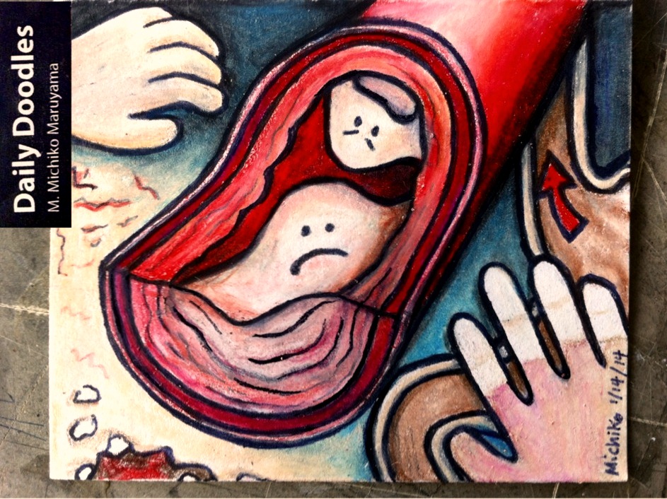
An article by F. Wigley states that “Although scleroderma is generally considered a fibrosing disease of the tissues, it is now recognized that the underlying vascular disease is playing a fundamental role in its pathogenesis and associated tissue injury” [3]. The vascular changes seen in systemic sclerosis include hyperplasia of the intima (inner layer) and narrowing of the lumen as illustrated in the center of the doodle with the cross-section of the blood vessel.
Scleroderma can be divided into two main forms: systemic and localized. Systemic scleroderma can be further divided into limited and diffuse. It is the diffuse form of scleroderma that involves the kidney, lung and heart.
In limited scleroderma, skin involvement is limited to the face, neck and distal extremities. The drawings around the edge of the Daily Doodle illustrate the CREST Syndrome, a variant of Limited Scleroderma.
CREST Syndrome:
- C – Calcinosis (bottom left of doodle)
- R – Raynaud’s phenomenon (bottom right of doodle)
- E – Esophageal motility dysfunction (right side of doodle)
- S – Sclerodactyly of the fingers (top left of doodle)
- T – Telangiectaes (top left of doodle)
Calcinosis cutis occurs when calcium is deposited in the skin and soft tissues. The deposits can vary in size and can be visualized on plain x-ray. They frequently occur in the finger pads, palms, extensor surfaces of the forearm, olecranon and prepatellar bursae. They usually present as persistent firm, nontender lumps. Occasionally, they may ulcerate through the skin producing a chalky white drainage.
Raynaud’s phenomenon, shown in the bottom right hand corner of the doodle, is a phenomenon when cold temperature or stress causes the fingers to blanche. If prolonged, they may become cyanotic, appearing red and dusky. Raynaud’s Phenomenon is caused by vasospasm and thickening of vessel walls in the digits. It can lead to ischemia of the digits, ulceration and gangrene. To treat Raynaud’s phenomenon, keep hands warm! Avoid the cold and if the patient smokes cigarettes, encourage and help them to quit. If symptoms are severe, consider calcium-channel blockers.
Sclerodactyly refers to a clawlike appearance of the hand caused by tightening of the skin.
References
- S. Agabegi and E. Agabegi. “Step-Up to Medicine”. Third Edition. Lippincott Williams and Wilkins. 2013
- Scleroderma Society of Canada: www.scleroderma.ca
- F. Wigley. “Vascular Disease in Scleroderma”. PubMed. 2009. www.ncbi.nlm.nih.gov/pubmed/19067252
- Fauci, Braunwald, Kasper, Hauser, Longo, Jameson, Loscalzo. Harrison’s Principles of Internal Medicine. 17th Edition. McGraw Hill Medical. 2008.
“Scleroderma – CREST Syndrome” by Michiko Maruyama